Diseases & Conditions
Rheumatoid Arthritis
Rheumatoid arthritis (RA) is one type of inflammatory arthritis. It is a chronic disease that can cause pain and stiffness in multiple joints throughout the body. It can affect any joint, but most often starts in the small joints of the hands and feet. In many cases, rheumatoid arthritis will affect joints on both sides of the body — both knees or both hands, for example.
According to the Centers for Disease Control and Prevention, approximately 1.5 million adults in the U.S. are living with rheumatoid arthritis, and 71 out of 100,000 people are diagnosed with RA every year.
Women are 2 to 3 times more likely to get RA than men. And although symptoms most commonly develop between the ages of 30 and 60, younger people can also be affected.
Children and adolescents with similar symptoms are often diagnosed with juvenile arthritis, which is also an autoimmune disease. There are several types of juvenile arthritis; however, nearly all of them are different from rheumatoid arthritis in adults. This is why the term "juvenile rheumatoid arthritis (JRA)" is no longer widely used and is known as juvenile idiopathic arthritis (JIA) instead.
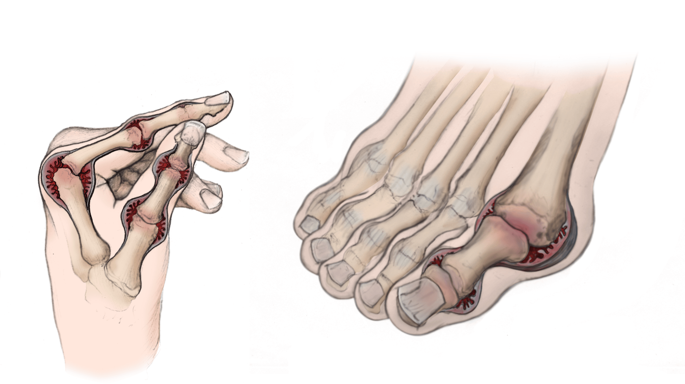
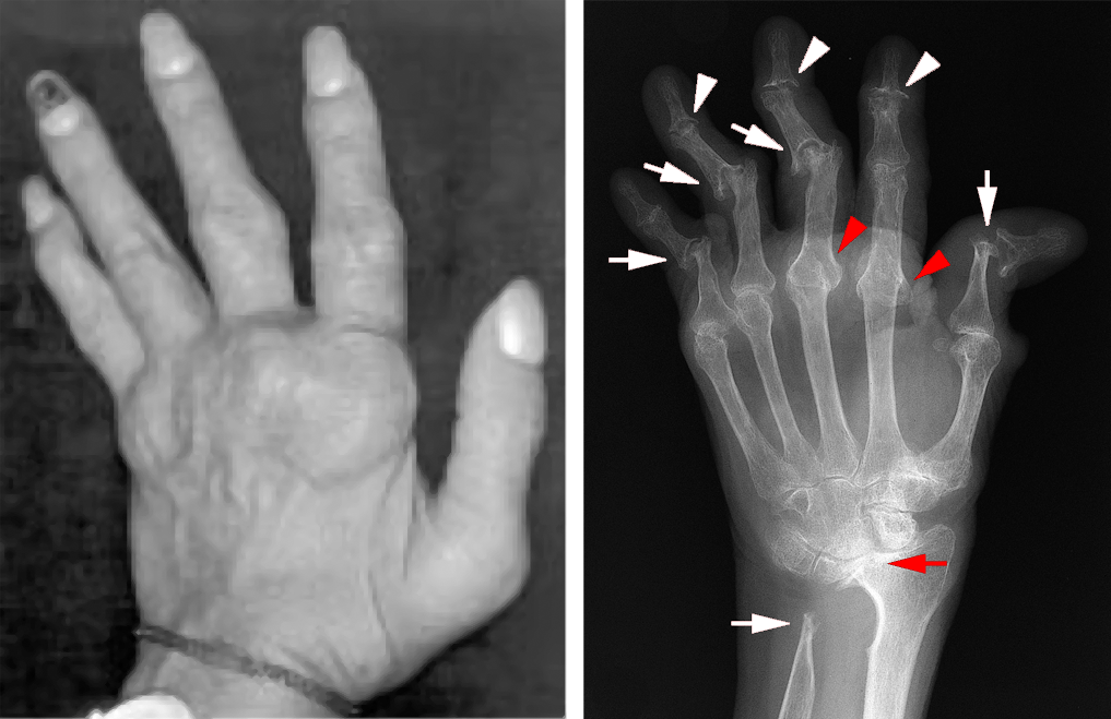
Over time, untreated rheumatoid arthritis causes joint damage and may result in serious disability. Fortunately, new medications now frequently prevent the progression of joint damage. The severe joint deformity often associated with RA — such as bent, crooked fingers and knuckles — is becoming uncommon due to these advances in treatment.
Although there is no cure for RA, early diagnosis and proper treatment can usually prevent joint destruction and other complications.
For joints that are severely affected by rheumatoid arthritis despite medical management, there are orthopaedic procedures that can greatly improve function, mobility, and overall quality of life.
Anatomy
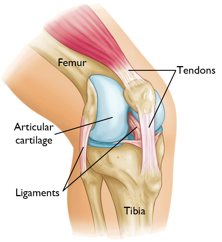
A joint is where the ends of two or more bones meet. There are different types of joints in the body — the most common type is a synovial joint. These types of joints — such as the knee, hip, and shoulder — are structured to make movement possible.
In a synovial joint, strong connective tissues called ligaments and tendons connect the bones to each other and to the surrounding muscles that stabilize the joint. The ends of bones are covered with a cushion of articular cartilage, a slippery tissue that helps the bones glide easily across each other during movement.
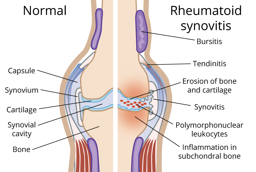
Synovial joints are surrounded with a thin lining of tissue — called synovium. The synovium produces hyaluronic acid and other substances that lubricate the joint and makes it easier to move.
Tendons that run through tight tunnels (called tendon sheaths) also have synovium around them to help them glide within the tunnels. These sheaths are found in the fingers, wrists, ankles, and toes. The synovial tissue within tendon sheaths is called tenosynovium. Rheumatoid arthritis can sometimes affect tenosynovium and cause swelling in the tendon sheaths. When this happens, the tenosynovial tissue can invade and damage the tendons, and the tendons may rupture (tear partially or completely).
Description
Rheumatoid arthritis (RA) is an autoimmune disease. This means that instead of protecting the body from infection, certain cells within the immune system begin attacking normal tissue. In the case of RA, immune cells attack the synovial tissue that lines the joints and/or the tenosynovium inside tendon sheaths.
How It Happens
In RA, immune cells release substances that inflame the synovium, causing it to thicken and swell. As the disease progresses, the abnormal synovium invades surrounding tissues, and produces chemical substances that destroy the cartilage and bone of the joint surface.
The ligaments, tendons, and muscles that support the joint can also be damaged. Tenosynovium can also be involved with the inflammation caused by rheumatoid arthritis. The tenosynovial tissue can invade and damage tendons, and the tendons may rupture (tear). This often occurs in the hands. Sometimes the first sign of rheumatoid arthritis is swelling and inflammation of the tendon compartments in the hand.
Weakened ligaments can lead to:
- Joint deformity
- Contractures (a permanent tightening that causes the joint to shorten and stiffen)
- Increased pain
Rheumatoid arthritis can also cause damage to blood vessels, skin, lungs, eyes, heart, and the nervous system. It can lead to low bone mass and osteoporosis.
Early diagnosis and treatment of RA can help control the disease and prevent lasting damage to joints and other structures.
Cause
The exact cause of RA is not known. It is not an inherited disease; however, researchers believe that some people have genes that make them vulnerable to the disease. Doctors suspect that it takes a chemical or environmental "trigger" to activate the disease in people who carry the genes. When the body is exposed to this trigger, the immune system responds incorrectly. Infection and smoking are two likely triggers.
Symptoms
RA affects people differently, with symptoms that can range from mild to severe. Many people experience ongoing mild symptoms throughout their lives, with occasional flares of more painful symptoms.
The most common rheumatoid symptoms are:
- Pain
- Fatigue
- Stiffness, especially morning stiffness that may last hours
- Swelling in more than one joint; the joints can ache even at rest
Other rheumatoid symptoms include:
- A feeling of warmth around the joint
- Symptoms throughout the body, such as fever, loss of appetite, and decreased energy
- Weakness due to a low red blood cell count (anemia)
- Nodules, or lumps, especially around the elbow
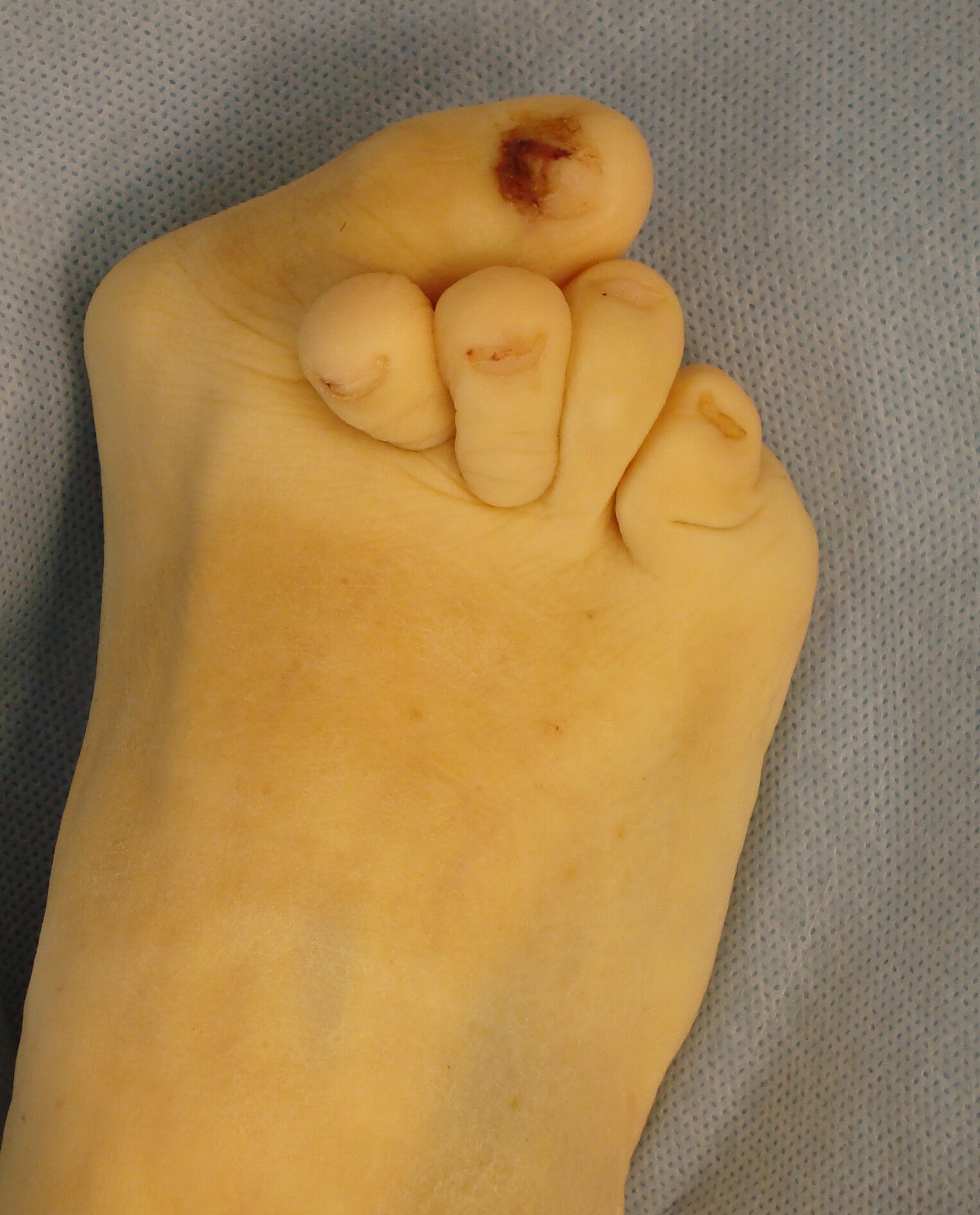
- Deformities and contractures of the joint (with long-lasting disease)
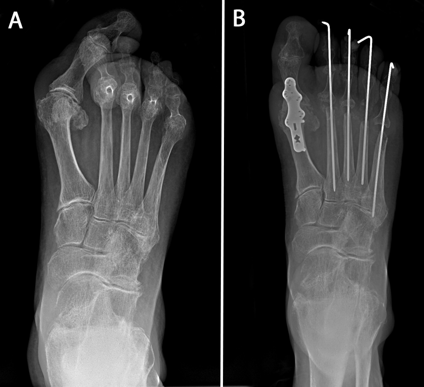
- Foot pain, bunions, and hammer toes (with long-lasting disease)
Patients with severe rheumatoid arthritis typically have multiple affected joints in the hands, arms, legs, and feet. Joints of the neck may be involved as well.
Doctor Examination
There is no single test or finding that confirms rheumatoid arthritis, so doctors use results from a patient’s health history, a physical examination, and several laboratory tests to rule out other conditions and make a diagnosis.
Medical History
Because symptoms typically develop slowly over time, rheumatoid arthritis can be difficult to diagnose in the early stages.
As part of the office visit, your doctor will take a complete medical history, including asking you about:
- Your general health
- Medications you take
- Your joint pain and other symptoms – when they started and whether the symptoms have changed over time
- Whether you or any family member has a history of arthritis or any autoimmune disorders
Physical Examination
During the physical examination, your doctor will look for:
- Signs of RA, including swollen or tender joints, limitation of joint motions, and early deformity.
- Signs of inflamed tendon sheaths in the hands or wrists
- Tendon ruptures (complete or partial tears)
The doctor will check both sides of the body because rheumatoid arthritis is often present bilaterally (on both sides).
Laboratory Tests
Certain blood tests can reveal signs of RA:
- Rheumatoid factor, an antibody that is present in about 85% of people with RA.
- Anti-CCP antibodies. These antibodies to cyclic citrullinated peptide/protein are found in many people with RA and are more specific to RA than rheumatoid factor.
- Erythrocyte sedimentation rate (ESR or "sed" rate) or C-reactive protein (CRP) These are common tests used to measure inflammation in the body. ESR and/or CRP are usually elevated in people with RA.
When analyzed all together, these blood tests are very useful in making a diagnosis of RA. It is important to note that it is possible to have normal bloodwork (negative rheumatoid factor) and still have rheumatoid arthritis. When this occurs, it is called seronegative rheumatoid arthritis.
Imaging Tests
X-rays. X-rays create images of dense structures like bone. Because bone and joint damage occur later in the course of the disease, X-rays may not be very useful in detecting RA in the early stages, although soft tissue swelling around a joint can be seen on a regular X-ray.
Your doctor may, however, use X-rays to help rule out other possible diagnoses. And if you have RA, your doctor may use periodic X-rays to help monitor any progression of the disease.
Criteria for Diagnosing Early-Stage Rheumatoid Arthritis
To help prevent RA from progressing to joint damage, the American College of Rheumatology has developed specific, comprehensive criteria to guide doctors in diagnosing RA in earlier stages of the disease.
In general, a positive diagnosis of RA can be made if the following criteria are met:
- Inflammatory arthritis in three or more joints
- Arthritis symptoms that have lasted for at least 6 weeks
- Positive serological test: rheumatoid factor present in blood testing and/or positive anti-CCP
- Elevated erythrocyte sedimentation rate (ESR or "sed" rate) or elevated C-reactive protein (CRP)
- Other possible causes of symptoms have been ruled out
If your doctor suspects RA, they may refer you to a rheumatologist. Although your symptoms and the results from a physical examination and tests may be consistent with RA, a rheumatologist will be able to determine the specific diagnosis. There are other less common types of inflammatory arthritis that should be considered.
Treatment
Although there is no cure for rheumatoid arthritis, there are many treatment options that can help relieve joint pain and improve functioning.
Medical management is key to prevention progressive disease, but surgical management is usually necessary when a person has painful joint destruction and/or tendon ruptures.
Rheumatoid arthritis is often treated by a team of healthcare professionals. These professionals may include family doctors, orthopaedic surgeons, rheumatologists, physical and occupational therapists, social workers, and rehabilitation specialists.
Medications and Medical Management
Medications used to control rheumatoid arthritis fall into two categories:
- Those that relieve symptoms
- Those that can modify the course of the disease (DMARDs), which are usually managed by a rheumatologist
Often, symptom relief medications and DMARDs are used together to decrease the doses of the stronger drugs, as well as to supplement the treatment plan during flare-ups of rheumatoid arthritis. The orthopaedic surgeon or general practitioner may start symptom relieving medications until the patient has been assessed and referred to a rheumatologist
Researchers are also working on biologic agents that can interrupt the progress of the disease. These agents target specific chemicals in the body to prevent them from acting on the joints.
Medications for Symptom Relief in Rheumatoid Arthritis
Non-steroidal anti-inflammatory drugs (NSAIDs). Anti-inflammatory drugs like naproxen and ibuprofen may relieve pain and help reduce inflammation. NSAIDs are available in both over-the-counter and prescription forms.
Corticosteroids. Medications like prednisone are potent anti-inflammatories. They can be taken by mouth (pills) or injected into an inflamed joint or tendon sheath. Due to the side effects associated with extended use of corticosteroids, they are generally prescribed on a short-term basis.
Although treatment with NSAIDs or short-term corticosteroids may relieve symptoms, it will not stop the progression of the disease. Specific medicines called disease-modifying anti-rheumatic drugs (DMARDs) are designed to stop the immune system from destroying the joints. The appropriate use of these medications is directed by a rheumatologist.
Disease-Modifying Anti-Rheumatic Drugs (DMARDs)
Disease-modifying anti-rheumatic drugs (DMARDs) help to slow the progress of RA by decreasing the body's overactive immune response. They reduce inflammation, prevent joint damage, and relieve painful symptoms.
There are two types of DMARDS:
- Conventional nonbiologic types
- Newer biologic agents
While both types target the immune system, biologics target specific immune cell types.
Nonbiologic DMARDS. There are many nonbiologic DMARD options for treating RA. They are typically in pill form, sometimes taken just once a week.
These conventional DMARDs do not provide immediate symptom relief — it can take weeks to months before you can tell whether the drug is working for you. In the meantime, your doctor may continue to prescribe a faster-acting nonsteroidal anti-inflammatory medication or a corticosteroid, such as prednisone. The patient is typically weaned (gradually takes less and less over time) off of anti-inflammatories once the DMARD has had a chance to work.
More than one DMARD can be used at the same time or added to your treatment plan if needed. Your doctor will monitor your symptoms and work with you over time to determine the most effective approach.
The most commonly used nonbiologic DMARDs are:
- Methotrexate
- Sulfasalazine
- Hydroxychloroquine
- Leflunomide
DMARDs used in patients whose disease does not respond to the initial medications include:
- Azathioprine
- Cyclosporine; however, these agents have fallen out of favor given the availability of newer, more effective agents (listed below)
Biologic DMARDs. Biologic DMARDs prevent joint damage by blocking the activation of specific immune cell types that stimulate inflammation. Different types of biologics target specific cells involved in the inflammatory process.
Although more expensive than conventional DMARDs, biologics act quickly to decrease the body's overactive immune and inflammatory activity. Their effect can be felt within 2 to 6 weeks of treatment.
In most cases, biologics are prescribed when arthritis symptoms persist after treatment with conventional DMARDs. For many patients, a combination of a conventional DMARD (most often methotrexate) and a biologic agent effectively manages symptoms and prevents joint damage.
Biologic agents are usually injected either subcutaneously (under the skin) or intravenously (through a tube inserted into a vein).
- Subcutaneous injections are typically done at home by the patient or a caregiver.
- Intravenous (IV) medications are given in the treating physician's office, at a hospital, or at an infusion center.
The most commonly used and effective biologics that are FDA-approved for treatment of RA are tumor necrosis factor inhibitors (anti-TNF). These medications decrease inflammation by blocking the tumor necrosis factor — a type of protein that activates inflammation during the immune response.
- Etanercept — given every week subcutaneously
- Adalimumab — given every 2 weeks subcutaneously
- Infliximab — given every 6 to 8 weeks intravenously
- Certolizumab pegol — given every 2 to 4 weeks subcutaneously
- Golimumab — given monthly subcutaneously
- Simponi ARIA — given every 8 weeks intravenously
There are other, less common biologic agents that can interrupt the inflammatory process, including:
- Tocilizumab (Actemra)
- Sarilumab (Kevzara)
- Abatacept (Orencia)
These too, are given either subcutaneously or through IV every few weeks.
Janus kinase (JAK) inhibitors. There is a newer group of DMARDS called JAK inhibitors. They include:
- Tofacitinib (Xeljanz)
- Upadacitinib (Rinvoq)
- Baricitinib (Olumiant)
These medications have the advantage of being in pill form.
Potential Side Effects of DMARDs and JAK Inhibitors
Each DMARD — conventional and biologic — has individual side effects. Examples of conventional DMARD side effects include:
- High blood pressure
- Liver damage
- Nausea
- Increased risk of serious infection; this is a major risk of treatment with any DMARD because DMARDs suppress the body's inflammatory response — which is the body’s normal method for fighting infections
Many patients who use biologics experience skin irritation at the injection site.
Along with the traditional risks of DMARDS, people taking JAK inhibitors have the additional risk of developing blood clots within blood vessels.
Patients require evaluation before starting DMARDs to rule out contraindications to the medications (meaning the patient should not take a certain medication due to the harm it would cause) and monitoring while using them to watch for serious side effects.
Patients are also screened for infections, including Hepatitis B and C and tuberculosis, before being prescribed these medications.
Talk to your doctor about all the medications included in your treatment plan to make sure you understand potential side effects and are prepared to quickly report them to your doctor should they occur.
Choosing the Right Medication for Your Rheumatoid Arthritis
Because there are now so many different medications available to treat rheumatoid arthritis, the decision about which medications to use depends on several factors, including:
- Disease severity
- Response to treatment
- Cost
- Side effects
- Other pre-existing conditions or medical problems
Physical Therapy and Exercise
Exercise is an important part of a treatment program. The physician and physical therapist may work with patients to develop an exercise program that helps strengthen the muscles around affected joints without stressing them.
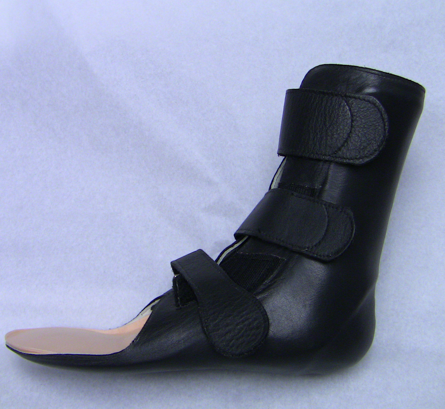
In some cases, your doctor may recommend upper extremity (arm) and/or lower (leg) extremity braces or splints for specific joints to help reduce stress on your joints and prevent deformity.
Surgical Treatment
Your doctor may recommend orthopaedic surgery depending upon the extent of cartilage damage and your response to nonsurgical options.
Synovectomy
In a synovectomy, the synovial joint lining damaged by rheumatoid arthritis is removed to reduce pain and swelling.
Synovectomy may be effective if the disease is limited to the joint lining and has not yet severely affected the articular cartilage that covers the bones. Generally, the procedure is used to treat only the early stages of RA. The synovitis can recur (come back) unless adequate medical management is ongoing. With the newer medical treatments available today, synovectomy is required less frequently.
Tendon Surgery
If tendons rupture, reconstructive surgery can restore function. Tendon repairs or tranfers can be very successful in the hand.
Trigger fingers and tenosynovitis in the hand and wrist can require:
- Partial release of tight tunnels
- Partial flexor tendon excision (removal) in the finger
- Tenosynovectomy to prevent tendon ruptures
Joint Rebalancing
In the early stages of rheumatoid arthritis, joints, especially the small joints of the hands and fingers, can become loose and unbalanced, causing deformities and contractures (when the tissue becomes stiff, constricted, and/or shortened) that interfere with function.
If the cartilage is still intact, a surgeon can perform procedures to release tight structures, transfer tendons, and tighten loose ligaments to rebalance the joint alignment and improve function. Joint rebalancing in larger joints is required during joint replacement to correct deformities and contractures.
Fusion
Fusion of the affected joints is the most common type of surgery performed for RA. Fusion takes the two bones that form a joint and fuses them together to make one bone. Fusions are mainly used in joints of the foot and hand because they can relieve symptoms while still allowing excellent function.
- During the surgery, the specific joint is exposed and the remaining damaged cartilage is removed from each side of the joint.
- The ends of the two bones are shaped to fit together tightly, then held together with screws or a combination of screws and plates. This prevents the bones from moving and allows blood vessels and new bone-forming cells to cross the fusion site.
- During the healing process, the body grows new bone between the bones, joining the two bones firmly together.
Fusion surgery eliminates motion in the joint, but patients often don't notice the lack of motion because the fusion makes the joint less painful and more stable, which improves function. Orthopaedic surgeons are trained to choose the position — the angle or alignment — of the fused joint that will provide the best function.
When joint replacement is possible, fusions are rarely done in the large joints of the elbow, shoulder, hip, and knee because of the importance of their mobility to good function. Ankle replacement has also become a viable option.
Joint Replacement Surgery
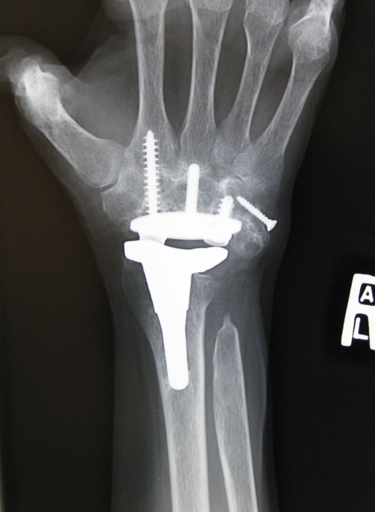
Joint replacement surgery is often effective at restoring painless joint movement. During this operation, your doctor removes the damaged cartilage and bone, and then positions new metal or plastic joint surfaces to restore the function of your joint.
Especially in the hand, a combination of specific small joint fusions and/or joint replacement improves function significantly. For the major joints, such as the elbow, shoulder, hip, and knee, these procedures can mean the difference between disability and an active life.
Preparing for Surgery
Many of the medications that help with RA also affect the ability of the body to heal wounds and fight infection. Ask your rheumatologist which medications you should stop taking for surgery and how soon to restart them. Your surgeon will also work with your rheumatologist or primary care doctor to review which of your medications will need to be stopped prior to surgery.
Patients who have rheumatoid arthritis in the cervical spine (neck) can have a dangerous laxity (looseness) in the ligaments around the first two cervical vertebrae. This can be seen with pre-operative X-rays. If you have this issue and are being given general anesthesia, your anesthesiologist will take special precautions when positioning your head and neck.
As with any orthopaedic surgery, smoking is a risk factor for post-operative infection. It is very important to work with your doctors to help you stop or decrease your use of nicotine before surgery. Learn more: Surgery and Smoking
Outcomes
Rheumatoid arthritis can cause a wide range of disabling symptoms. Today, new medications may prevent progression of disease and joint destruction. Early diagnosis and treatment can help preserve the joints.
In cases that progress to severe joint damage, surgery can help relieve your pain, increase motion, and help you get back to enjoying everyday activities.
Contributed and/or Updated by
Peer-Reviewed by
AAOS does not endorse any treatments, procedures, products, or physicians referenced herein. This information is provided as an educational service and is not intended to serve as medical advice. Anyone seeking specific orthopaedic advice or assistance should consult his or her orthopaedic surgeon, or locate one in your area through the AAOS Find an Orthopaedist program on this website.


