Diseases & Conditions
Ulnar Collateral Ligament (UCL) Injury
This article was written and/or reviewed by a member of American Shoulder and Elbow Surgeons (ASES).
An injury to the ulnar collateral ligament (UCL) of the elbow most often occurs in overhead throwing athletes due to high repetitive stresses leading to an overuse injury. The incidence of injury to the ulnar collateral ligament is rising in the U.S., particularly among amateur athletes.
While UCL injuries commonly occur in pitchers, they can occur in any individual who participates in repetitive overhead throwing or an activity that places stress on the elbow. This includes some track and field athletes, gymnasts, volleyball players, and position baseball and softball athletes.
Anatomy
The elbow joint is made up of three bones:
- The humerus (upper arm bone)
- The radius and ulna (the two bones of the forearm)
The elbow is a combination of a hinge joint which allows the elbow to bend (flex) and strengthen (extend); and a pivot joint that allows it to rotate so you can bring your palm up and down.
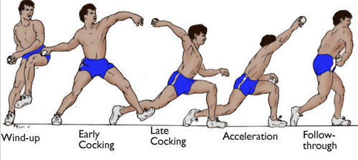
Since the humerus fits perfectly into the forearm, the elbow functions as a very stable joint for most daily activities. However, the throwing motion requires the elbow to bend during the cocking phase of throwing in a manner that distracts, or opens up, the inside part of the joint.
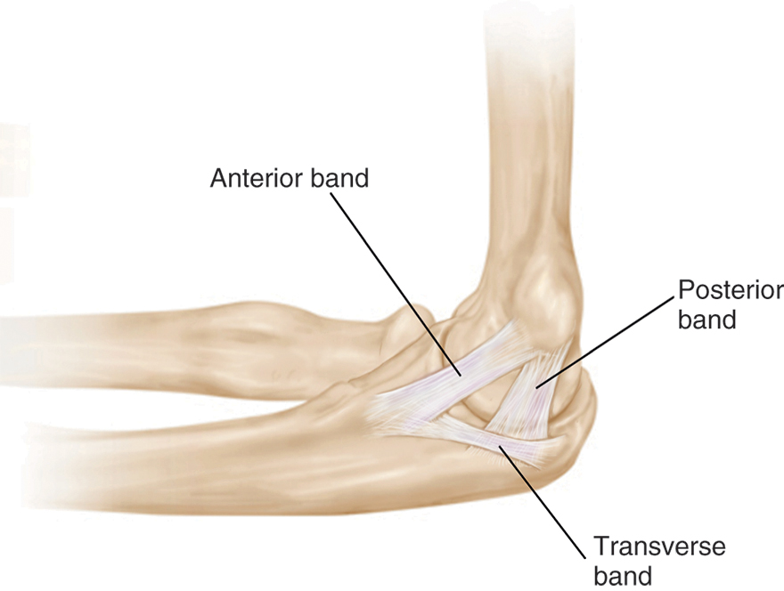
The ulnar collateral ligament is a thick band of tissue on the inside part of the elbow that is meant to resist the strong forces involved in overhead throwing. The ligament, which connects the humerus to the ulna, is actually made up of three separate, connected bands:
- The anterior (front) band. This is the most important structure in providing stability to overhead throwing athletes.
- The posterior (back) band.
- The transverse band, which connects the other two bands.
Cause
There are two main causes of ulnar collateral ligament injuries: injury and wear (degeneration).
Acute Tear
Some individuals and athletes may hear a “pop” or feel a sudden “snap” in their elbow with immediate pain. In this type of tear, the athlete will be unable to continue throwing or performing overhead activities with the injured arm.
The ulnar collateral ligament can also be injured during an acute elbow dislocation that occurs from a high energy fall onto an outstretched hand.
Overuse Tear
Most tears are the result of a wearing down of the ligament that occurs slowly over time.
- With repetitive valgus force on the elbow, which is the type of bending that occurs particularly in the late cocking phase of throwing, there is repetitive microtrauma to the ulnar collateral ligament.
- Over time, an individual may notice a gradual decline in their ability to compete at the same level. For instance, baseball pitchers often complain of a loss of velocity in their pitches, accompanied by loss of control and accuracy.
The type of bending that is required to throw a baseball, softball, or javelin is not a natural force for the elbow. As a result, it is important to maintain good throwing mechanics, strong forearm musculature, hip and shoulder flexibility, and good core strength to protect the elbow throughout a throwing maneuver. When this kinetic chain is disrupted, the ulnar collateral ligament experiences more stress and becomes more at risk of injury.
Iatrogenic Tear
Some surgeries may lead to unintentional injury to the ulnar collateral ligaments. In cases of valgus extension overload, individuals may develop bone spurs on the olecranon (bony tip of the elbow). Surgery to remove these bone spurs may lead to injury to the ligament, resulting in symptoms.
Symptoms
The most common symptoms of an ulnar collateral ligament tear include:
- Pain on the inside part of the elbow during throwing and overhead activities, or after a throwing session
- Inability to compete at the same level as before
- Loss of velocity during throws
- Loss of accuracy during throws
- Occasionally, tingling in the pinky and ring fingers
Tears that happen suddenly, such as from a fall or during a dislocation, usually cause intense pain.
Tears that develop slowly due to overuse may also cause pain and dysfunction.
- At first, the pain may be mild and occur only when throwing many pitches or at a high velocity.
- The pain may become more noticeable at rest and not improve with simple remedies such as rest, ice, and/or over-the-counter medication (e.g., aspirin, ibuprofen, or naproxen).
Since these injuries typically occur in athletes from overuse, it is important to discuss any symptoms, even if mild, with a coach, trainer, parent, and/or a medical professional immediately. Early identification of a problem — and taking prompt steps to address it — may help to prevent a more significant injury.
Doctor Examination
Medical History and Physical Examination
After discussing your symptoms and medical history, as well as reviewing your competitive schedule and goals for continuing to compete, your doctor will examine your elbow. The will:
- Check for any areas that are tender to the touch. Specifically, they will push on the inside of the elbow where the ligaments attach to the bone.
- Have you move your arm in several different directions to measure the range of motion of your elbow and shoulder.
- Test the strength of your elbow.
- Perform special tests to stress the ulnar collateral ligament, similar to the positions of throwing. These include the moving valgus stress test and the milking maneuver.
- Test the strength and sensation in your hand on the side of your injured elbow, as well as tap on the funny bone nerve (ulnar nerve). Since the ulnar collateral ligament is next to the nerve, you may have some symptoms or irritation related to it, which are important to identify.
Imaging Tests
Imaging tests are very helpful to diagnose an ulnar collateral ligament tear, as well as to rule out any other injuries.
- X-rays. The first imaging tests performed are usually X-rays. Because X-rays do not show the soft tissues of your elbow like the collateral ligaments, plain X-rays may be normal; they may, however, show some calcium build-up in the area of the ulnar collateral ligament or reveal small bone spurs.
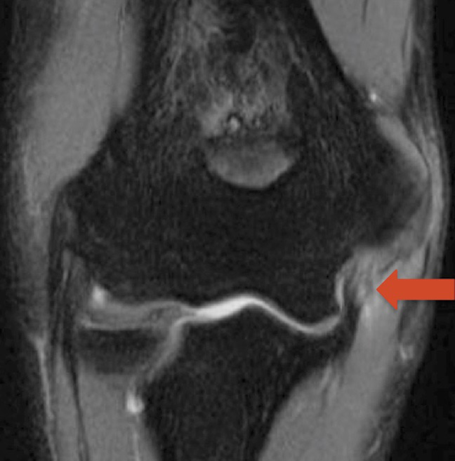
- Magnetic resonance imaging (MRI) scans. An MRI can better show the soft tissues, like the ulnar collateral ligament. Often, an MRI arthrogram is ordered, in which a special dye is injected into your elbow joint to help the doctor better see ligament tears, including small partial tears. An MRI can also show if the ligament is torn from the arm bone (humerus), from the forearm bone (ulna), or at the middle of the tendon. When the tear occurs at the ulna, it is less likely to heal on its own.
Treatment
When determining treatment for an ulnar collateral ligament tear, especially if it is an overuse injury in a throwing athlete, it is important to determine the athlete's goals.
The goal of any treatment is to reduce pain and restore function. Specifically, your doctor will consider:
- Your age
- Your activity level
- Your desire to continue with competitive throwing, especially pitching.
- Your goals to compete at higher levels of competition
- Your willingness to change positions; for instance, playing first base places less stress on the elbow than playing an outfield position, catching, or pitching
- The current point of time in your sport's season (i.e., offseason, preseason, playoffs)
Nonsurgical Treatment
Nonsurgical treatment options may include:
- Your doctor may suggest rest, ice, and elevation. Rest involves stopping all overhead and throwing activities.
- Nonsteroidal anti-inflammatory drugs (NSAIDs). Anti-inflammatory drugs like ibuprofen, aspirin, and naproxen can reduce pain and swelling.
- Steroid injections are typically not recommended by most surgeons for ulnar collateral ligament tears, especially partial tears, because the cortisone (steroid) may affect the soft tissue and lead to a larger tear and/or prevent healing. Some doctors or providers may recommend a platelet-rich plasma (PRP) injection. This is a type of injection where blood is drawn from you, and a special instrument is used to extract the plasma and platelets that have healing potential. Although current studies are inconclusive, early data is promising, and the evidence to support the use of PRP for UCL tears is growing.
After stopping throwing for about 6 to 12 weeks, your doctor will re-examine your elbow.
- If there is no more tenderness over the ligament and the special tests for the ulnar collateral ligament are not painful, the doctor will order a physical therapy program to work on throwing mechanics, and strengthening your forearms and your core muscles, before starting you on a throwing program.
- As you work through a specific throwing program, if your velocity and control improve without any pain, you will be cleared for full competition.
Nonsurgical treatment avoids the risks of surgery. However, it does limit activities and may only delay the inevitable need for surgery, which has a much longer rehabilitation program.
Surgical Treatment
Reconstruction (Tommy John Surgery). Surgery for an injured ulnar collateral ligament was historically career-ending, especially for baseball pitchers.
Then, in 1974, Dr. Frank Jobe developed a procedure that was performed on a major league pitcher named Tommy John. Since that time, the surgical procedure that is commonly known as a "Tommy John," UCL reconstruction, has become the gold standard, enabling a great many athletes at all levels to return to play.
In this procedure, your surgeon will reconstruct (or rebuild) your ulnar collateral ligament using a tissue graft, which acts as a scaffold for a new ligament to grow on. Typically, the graft is one of the patient's own tendons, either:
- An extra tendon in the wrist called the palmaris longus
- For people who are born without a palmaris longus (as a percentage of the general population is), one of the hamstring tendons in the knee
Another option is to use an allograft — meaning a tendon from a cadaver. This is typically avoided in competitive athletes, as there has been a reported lower return to sport with this option.
Reconstruction surgery is performed with either general anesthesia or regional anesthesia with sedation. Your surgeon and anesthesiologist will discuss the options with you prior to your procedure.
For the procedure, the surgeon makes an incision on the inside part of your elbow. After harvesting the tendon graft to be used, the surgeon will usually drill holes in the arm bone (humerus) and forearm bone (ulna) to secure the graft and stabilize the elbow. Sometimes, an implant, such as a plastic anchor, may be used in addition to the graft. If a portion of your own ulnar collateral ligament is intact (not torn), your surgeon may incorporate that into the procedure.
Repair. In recent years, some surgeons have begun repairing the injured ligament rather than rebuilding the ligament with a graft. This typically involves using a plastic anchor and a strong suture to repair and reinforce your own ligament back to the bone.
The benefit of repair over reconstruction is that the rehabilitation protocol is significantly shorter. Repair may be an option for you based on:
- Your sport
- Your position
- Your tear type
- The quality of your elbow tissues
Discuss with your surgeon whether you are a candidate for repair.
Recovery
After surgery, you will feel pain. This is a natural part of the healing process. Your doctor and nurses will work to reduce your pain.
Medications are often prescribed for short-term pain relief after surgery. Many types of medicines are available to help manage pain, including opioids, non-steroidal anti-inflammatory drugs (NSAIDs), and local anesthetics. Your doctor may use a combination of these medications to improve pain relief, as well as minimize the need for opioids.
Be aware that although opioids help relieve pain after surgery, their use has risks and complications. These medications can be addictive and potentially dangerous. It is therefore important to use opioids only as directed by your doctor, to use as little as possible for as short a time as possible, and to stop taking them as soon as your pain starts to improve. Tell your doctor if your pain has not begun to improve within a few days after surgery.
Rehabilitation
Rehabilitation after surgery for an ulnar collateral ligament tear is critical to getting you back to your desired level of competition.
- Your surgeon will prescribe a specific rehabilitation protocol for you to follow. It is very important that you, your therapist, and your trainer follow this protocol carefully.
- Typically, after surgery there is a period of immobilization in a splint. Afterward, you may be placed into a hinged elbow brace that allows some motion but still protects the ligament repair or reconstruction that was performed.
- Your physical therapist and trainer will also work on your forearm strengthening, your core strengthening, and your hip mechanics. Once your range of motion has been optimized, you may start what is called a "Thrower's Ten Program."
- Specifically for baseball players, an interval throwing program will be initiated when your surgeon allows. This will control how many throws you make, the distance you throw, and how hard you throw at various intervals. It is important to follow this program thoroughly to avoid risk of early re-injury.
Following a reconstruction, expect a complete recovery to take about 12 to 18 months.
Outcome
In recent years, there has been a lot of research on the outcomes of ulnar collateral ligament reconstruction (Tommy John surgery). The ability of patients to return to sport varies; however, in most studies, about 80% are able to compete again at the same level as before the injury.
In those who are unable to return to play and undergo a second Tommy John surgery, the rates of success and return to sport are much lower.
Complications
After ulnar collateral ligament surgery, a small percentage of patients experience complications. In addition to the risks of surgery in general, such as blood loss or problems related to anesthesia, complications of ulnar collateral ligament surgery may include:
- Nerve injury. This typically involves the funny bone nerve (ulnar nerve), causing tingling in the pinky and ringer finger that often goes away on their own in time. Another small nerve crosses the arm at the lower part of the incision and provides sensation to the inside part of the forearm. If injured, you may experience some tingling, burning, or pain on the inner forearm.
- Infection. Patients are given antibiotics during the procedure to lessen the risk for infection. If an infection develops, additional surgery and/or prolonged antibiotic treatment may be needed.
- Fracture. Surgery involves drilling bone tunnels, which can make the bone fragile and lead to a fracture. This may require additional surgery for fixation of the bone fragments.
- Stiffness. Early range of motion in a hinged elbow brace is encouraged to avoid stiffness, but stiffness can still occur after surgery. Elbows are also prone to developing extra bone where it does not belong. This is called heterotopic ossification. If this occurs, you may require more surgery.
Contributed and/or Updated by
Peer-Reviewed by
AAOS does not endorse any treatments, procedures, products, or physicians referenced herein. This information is provided as an educational service and is not intended to serve as medical advice. Anyone seeking specific orthopaedic advice or assistance should consult his or her orthopaedic surgeon, or locate one in your area through the AAOS Find an Orthopaedist program on this website.


