Treatment
X-rays, CT Scans, and MRI Scans
Diagnostic imaging techniques help narrow the causes of an injury or illness and ensure that the diagnosis is accurate. These techniques include X-rays, computed tomography (CT) scans, and magnetic resonance imaging (MRI) scans.
These imaging tools let your doctor see inside your body to get a picture of your bones, organs, muscles, tendons, nerves, and cartilage. This is a way the doctor can determine if there are any problems.
X-ray
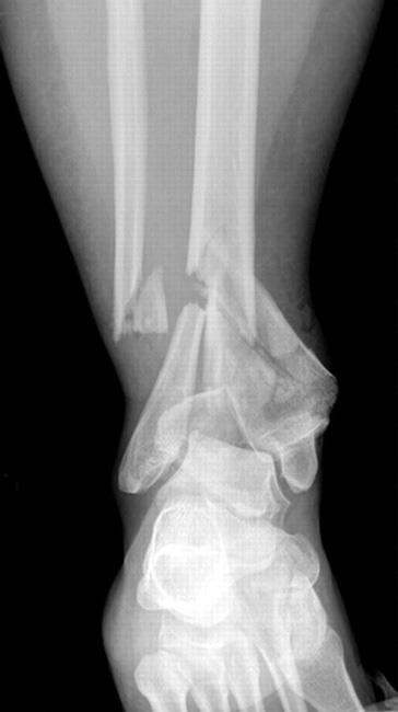
X-rays (radiographs) are the most common and widely available diagnostic imaging technique. Even if you also need more sophisticated tests, you will probably get an X-ray first, as X-rays can show some abnormalities better than the more sophisticated tests.
What Happens During an X-ray
The part of your body being pictured is positioned between the X-ray machine and photographic film or digital X-ray sensor. You have to hold still while the machine briefly sends electromagnetic waves (radiation) through your body, exposing the film to reflect your internal structure.
Bones, calcifications, some tumors, and other dense matter appear white or light because they absorb the radiation. Less dense soft tissues and breaks in bone let radiation pass through, making these parts look darker on the X-ray film.
You will probably be X-rayed from several angles. If you have a fracture in one limb, your doctor may want a comparison X-ray of your uninjured limb. Your X-ray session will probably be finished in about 10 minutes. The images are ready quickly. They are either developed from the X-ray film or sent as digital files to be viewed on a computer screen.
In some circumstances, a contrast material or dye may be injected into a joint while X-rays are taken. This procedure, which is called an arthrogram, helps to outline soft tissue structures in the joint. It may also confirm needle placement in the joint when fluid is removed or medication is injected into the joint.
X-rays may not show as much detail as an image produced with more sophisticated techniques. They are, however, the most common imaging tool used to evaluate an orthopaedic problem and are readily available in most doctors' offices and urgent care centers.
The level of radiation exposure from X-rays is not harmful, but your doctor will take special precautions if you are pregnant. Learn more: Pregnancy and X-rays
Computed Tomography (CT) Scan
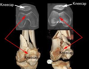
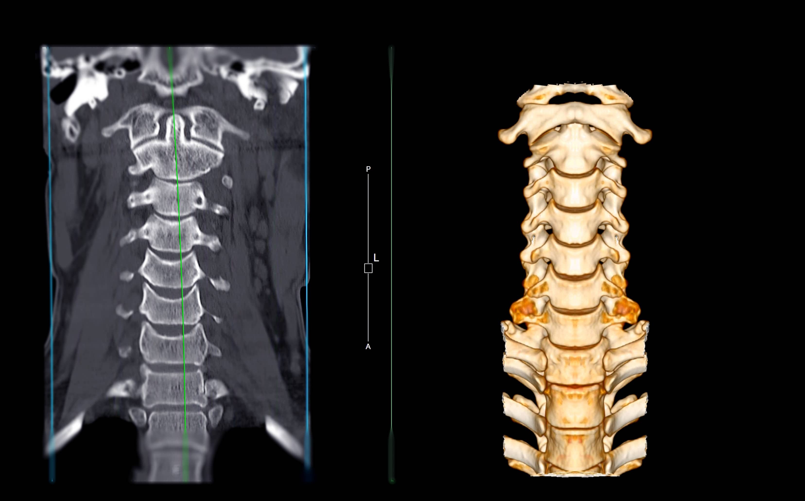
Computed tomography (CT) is an imaging tool that combines X-rays with computer technology to produce a more detailed, cross-sectional image of your body. A CT scan lets your doctor see the size, shape, and position of structures that are deep inside your body, such as organs, tissues, bone structures, or tumors.
You may need a CT scan if:
- You have a problem with a small, bony structure
- You have severe trauma to the brain, spinal cord, chest, abdomen, or pelvis
Tell your doctor if you are pregnant before undergoing a CT scan.
A CT scan costs more and takes more time than a regular X-ray. It can be done in either a hospital setting or an outpatient imaging center.
What Happens During a CT Scan
During the scan, you lie as motionless as possible on a table that slides into the center of the cylinder-like CT scanner. The process is painless. An X-ray tube slowly rotates around you, taking many pictures from all directions. A computer combines the images to produce a clear, two-dimensional view on a television screen.
Sometimes, you may be given a dye or contrast material to make certain parts of your body show up better.
Magnetic Resonance Imaging (MRI) Scan
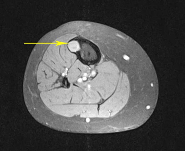
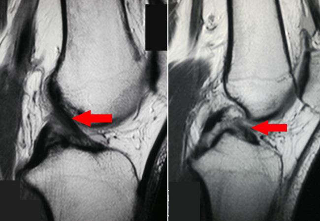
Magnetic resonance imaging (MRI) is another diagnostic imaging technique that produces cross-sectional images of your body. Unlike X-rays and CT scans, MRI scans work without radiation. The MRI tool uses magnetic fields and a sophisticated computer to take high-resolution pictures of your bones and soft tissues.
An MRI scan may be used to help diagnose a wide range of issues, including but not limited to:
- Torn knee ligaments
- Meniscus tears
- Rotator cuff tears
- Herniated disks
- Osteonecrosis
- Bone tumors
Tell your doctor if you have a pacemaker, implants, metal clips, or other metal objects in your body before you undergo an MRI scan. The MRI technologist and radiologist can determine if an MRI can be performed safely depending on which type of metal object is present.
Another somewhat recent development is the improved ability to scan a joint that has been replaced. Artificial joints contain a lot of metal, which can cause MRI scans to appear blurry, making it harder to see damage in the area around the implant. Some newer MRI machines have software, called metal artifact reduction sequences, or MARS, that reduces the blurring of the images. MARS may be used in certain patients to help to diagnose certain conditions. It does not take longer to perform this scan, and there is no additional risk for the patient.
What Happens During an MRI Scan
During the scan, you lie as motionless as possible on a table that slides into the tube-shaped MRI scanner. The MRI creates a magnetic field around you and then pulses radio waves to the area of your body to be pictured. The radio waves cause your tissues to resonate.
A computer records the rate at which your body's various parts (tendons, ligaments, nerves, etc.) give off these vibrations, and translates the data into a detailed, two-dimensional picture. You will not feel any pain while undergoing an MRI, but the machine may be noisy. The MRI technologist may offer you some ear protection (ear plugs or headphones) to help with the noise.
Another downside of MRI is that the tube can be very confining, and claustrophobic patients often have trouble holding still. Some patients require a one-time dose of medicine to help them relax enough to complete the scan.
It may take 30 to 60 minutes to do the study. Like a CT scan, an MRI scan may be done in a hospital or at an outpatient imaging center.
Other Imaging Tests
Other orthopaedic imaging tests include ultrasound and bone scan (nuclear imaging).
Ultrasound. Ultrasound uses high-frequency sound waves that echo off the body. It is painless, noninvasive, and does not require radiation. Ultrasound is used most often to look for blood clots, but can also show other problems, such as a Baker's cyst behind the knee or a rotator cuff tear in the shoulder.
Bone scan. A bone scan uses a small amount of radioactive material to identify areas of increased bone activity. The material is injected into a vein and is absorbed by areas that are forming new bone, such as a healing fracture, bone tumor, or bone infection.
The scan is done several hours after the injection. The radioactive material is eliminated quickly from the body.
A bone scan has the advantage of showing bone activity throughout the entire body.
Contributed and/or Updated by
Peer-Reviewed by
AAOS does not endorse any treatments, procedures, products, or physicians referenced herein. This information is provided as an educational service and is not intended to serve as medical advice. Anyone seeking specific orthopaedic advice or assistance should consult his or her orthopaedic surgeon, or locate one in your area through the AAOS Find an Orthopaedist program on this website.


