Diseases & Conditions
Vertical Talus
Vertical talus is a rare deformity of the foot that is diagnosed at birth. Because babies are born with the condition, it is also known as congenital vertical talus (CVT). It is one of the causes of a flatfoot in newborns. One foot, or both feet, may be affected.
Although it is not painful for the newborn or even the toddler, if it is left untreated, vertical talus can lead to serious disability and discomfort later in life.
Anatomy
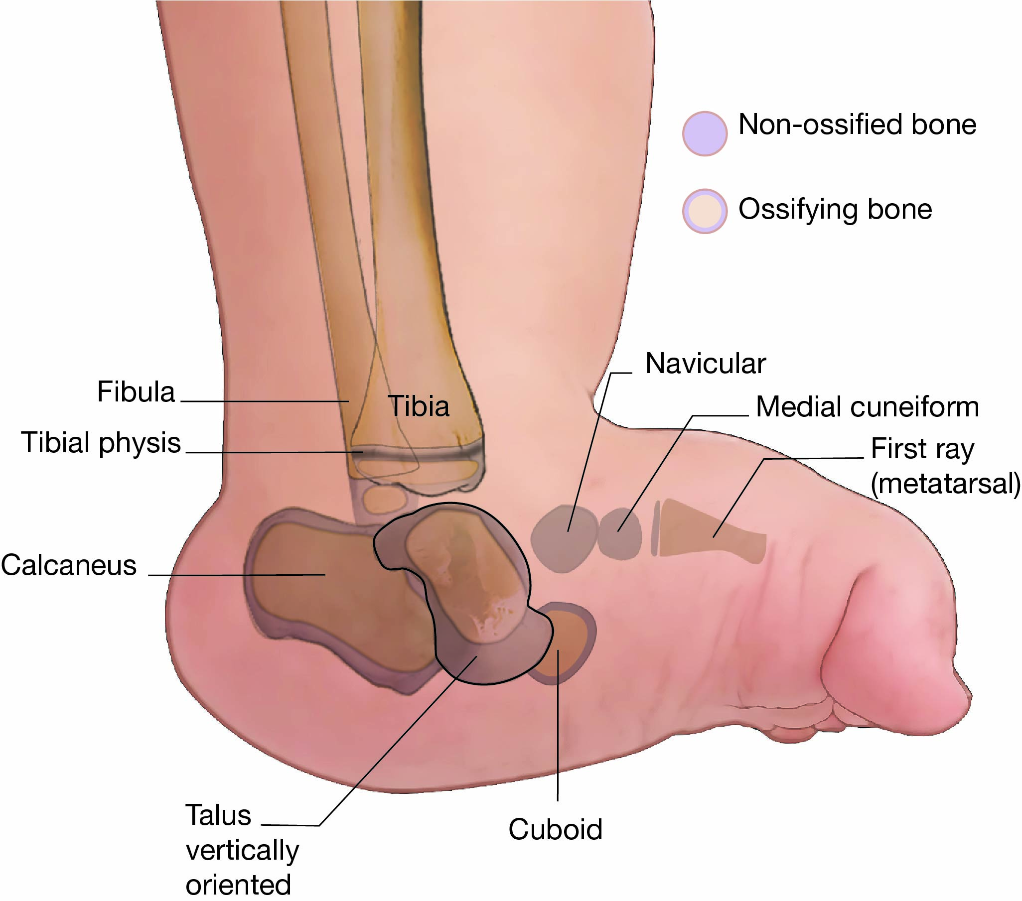
The talus (TAY-lus) is a small bone that sits between the heel bone (calcaneus) and the two bones of the lower leg (tibia and fibula). The tibia and fibula sit on top and around the sides of the talus to form the ankle joint. The talus is an important connector between the foot and the leg, helping to transfer weight across the ankle joint.
Description
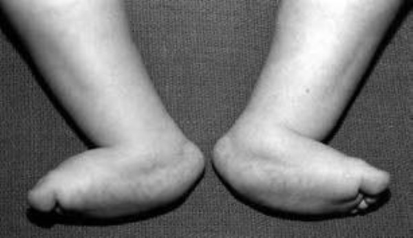
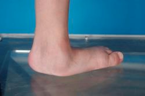
In vertical talus, the talus bone has formed in the wrong position, so the other bones in the foot are not lined up properly. As a result, the front of the foot points up and may even rest against the front of the shin. The bottom of the foot is stiff and has no arch. Instead, it usually curves out and is often described as "rocker bottom."
This is distinctly different than a clubfoot. Vertical talus is a rigid deformity, and the foot appears flat even when the child is not standing.
Vertical talus is usually diagnosed at birth. Other foot deformities in the newborn are more common, and vertical talus is often initially misdiagnosed as some other type of newborn flatfoot, such as an oblique talus or calcaneovalgus deformity. Some clinicians with less experience in these newborn foot conditions have even incorrectly thought of this as a clubfoot.
Cause
The exact cause of vertical talus in not known. Many cases, however, are associated with a neuromuscular disease or other disorder, such as arthrogryposis or spina bifida. Your child's doctor may decide to perform additional tests to discover whether your infant has any of these other conditions.
Doctor Examination
Pediatric orthopaedic doctors are able to diagnose vertical talus by simply examining the child. However, your child's doctor may order a special X-ray of your child's foot to confirm the diagnosis.
Treatment
The goal of treatment for vertical talus is to provide your child with a functional, stable, and pain-free foot.
It is important for vertical talus to be treated early. A vertical talus will not prevent your child from walking, but over time:
- Calluses and painful skin problems will develop.
- Finding shoes that fit properly will become challenging.
- Your child will start having pain that impacts their function.
Treatment usually involves a combination of nonsurgical and surgical intervention.
Nonsurgical Treatment
Initial treatment is usually nonsurgical and includes a series of stretching and casting designed to stretch tight tendons in the foot and bring the front part of the foot into a better alignment with the hindfoot (talus and calcaneus).
Surgical Treatment
If the initial nonsurgical treatment does not effectively correct the problem, your child may need a larger surgery as early as 9 to 12 months of age.
During the operation, your child's doctor will put the bones in the correct position and apply pins to keep them in place. Tendons and ligaments on the top part of the foot will be lengthened to allow the foot to be brought into a better position. The Achilles tendon also is lengthened as part of this procedure.
Recovery
After the operation, the doctor will apply a cast to your child's foot to keep it in the corrected position. Your child may stay in the hospital for at least 1 night after surgery to help control pain, and for the doctor to monitor any swelling in the foot.
After 4 to 6 weeks, the cast will be removed. A brace or special shoe is often used once the cast is removed to help prevent the deformity from returning.
Outcomes
Without treatment, your child's vertical talus will most likely result in future pain and disability.
With treatment, you can expect a stable and functional foot that should serve your child well throughout their life. The most important factor affecting your child's outcome is whether there is an underlying neuromuscular or genetic condition. If your child has no other conditions that limit function and development, you can expect your child to run and play without pain, and to wear normal shoes.
Your child's doctor will likely recommend repeat clinic visits over the years to observe the growth and development of your child's foot.
Contributed and/or Updated by
Peer-Reviewed by
AAOS does not endorse any treatments, procedures, products, or physicians referenced herein. This information is provided as an educational service and is not intended to serve as medical advice. Anyone seeking specific orthopaedic advice or assistance should consult his or her orthopaedic surgeon, or locate one in your area through the AAOS Find an Orthopaedist program on this website.


