Diseases & Conditions
Recurrent and Chronic Elbow Instability
This article was written and/or reviewed by a member of American Shoulder and Elbow Surgeons (ASES).
Elbow instability is a sense of looseness in the elbow joint that may cause the joint to catch, pop, or slide out of place during certain arm movements. It most often occurs as the result of an injury — typically, an elbow dislocation.
- A dislocation is an event when the joint stays out of place. This type of injury can damage the bone and ligaments that surround the elbow joint.
- The term subluxation refers to a partial dislocation, which means the joint partially slips out, but then goes back in place.
- When the elbow is loose and repeatedly feels as if it might slip out of place, it is called recurrent or chronic elbow instability.
Anatomy
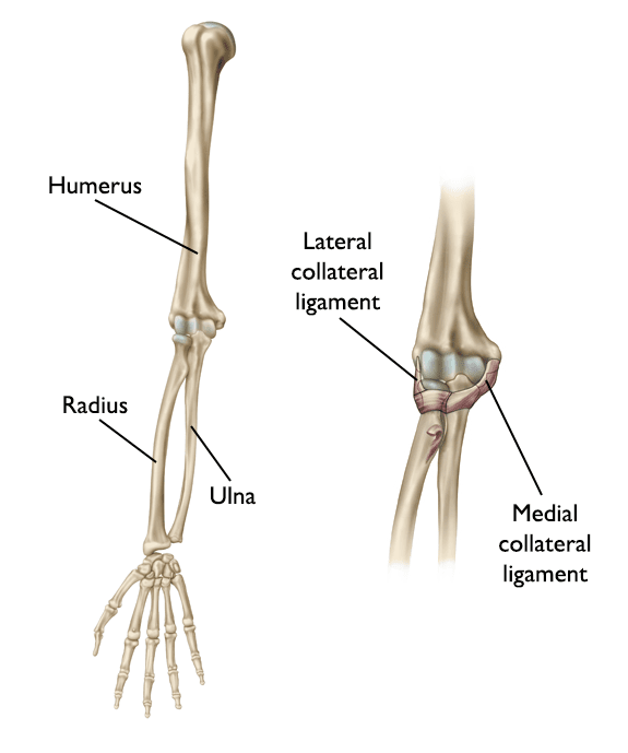
The elbow is made up of:
- The humerus (upper arm bone)
- The radius and ulna (the two bones in the forearm)
On the inner and outer sides of the elbow, strong ligaments (collateral ligaments) hold the elbow joint together and work to prevent dislocation/subluxation events. The two important ligaments are:
- The lateral (outside) collateral ligament
- The medial (inside) collateral ligament
The muscles that cross the elbow joint also contribute to the stability of the joint.
Description
There are three common types of recurrent elbow instability:
- Posterolateral rotatory instability. The elbow slides in and out of the joint due to an injury of the lateral collateral ligament complex. This is the most common type of recurrent elbow instability. Associated fractures can also occur with this type of instability.
- Valgus instability. The elbow is unstable due to an injury of the medial collateral ligament.
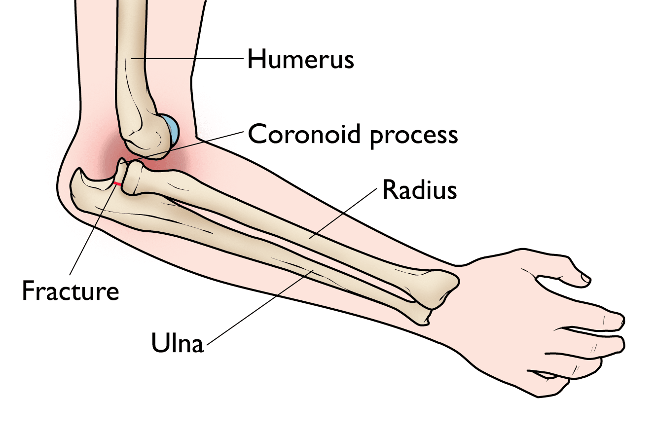
- Varus posteromedial rotatory instability. The elbow slides in and out of the joint due to an injury of the lateral collateral ligament complex, in addition to a fracture of the coronoid portion of the ulna bone on the inside of the elbow.
Cause
There are different causes for each of the different patterns of recurrent elbow instability:
- Posterolateral rotatory instability is typically caused by a trauma, such as a fall on an outstretched hand. It may also develop as a result of a previous surgery, or longstanding elbow deformity.
- Valgus instability is most often caused by repetitive stress as seen in overhead athletes (such as baseball pitchers). Like the other forms of recurrent elbow instability, it may also result from a traumatic event.
- Varus posteromedial rotatory instability is typically caused by a traumatic event, such as a fall.
Symptoms
- Recurrent elbow instability may cause pain, locking, catching, or clicking of the elbow.
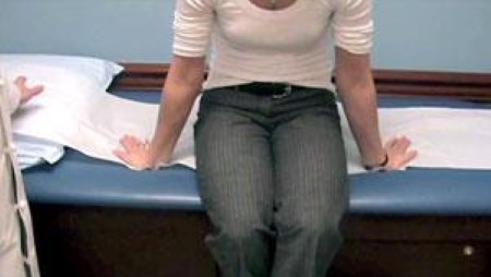
- You may also have a sense of the elbow feeling like it might pop out of place. This feeling commonly occurs while pushing off from a chair, specifically with posterolateral rotatory instability.
- When there is an injury to the medial collateral ligament, overhead athletes may feel pain on the inside of their elbow when throwing, or experience a loss in throwing velocity (speed) and ball control.
Doctor Examination
Medical History and Physical Examination
After discussing your symptoms and medical history, your doctor will examine your elbow. They will:
- Check to see whether the elbow is tender in any area
- Check to see if there is a deformity
- Have you move your arm in several different directions to test for instability or a popping or sliding sensation
- Test your arm strength
- Make sure there are no injuries to your nerves
Many cases of elbow instability can be diagnosed based solely on a medical history and physical examination results.
Imaging Tests
X-rays. Although X-rays cannot show soft tissues like ligaments, they can be useful in identifying fractures, dislocations, or subtle changes in elbow alignment.
Ultrasound. An ultrasound scan uses sound waves to capture pictures of your anatomy. This dynamic exam can show tears in ligament, muscles, or tendons. It may be used to create side-by-side images of the injured elbow and healthy elbow for your doctor to compare and identify any differences.
Magnetic resonance imaging (MRI). An MRI scan creates better images of soft tissues than an X-ray, and it may show tears in the ligaments, muscles, or tendons. MRI scans are not always necessary to diagnose elbow instability. However, they can be helpful in confirming the diagnosis, especially in overhead athletes where there is concern about the medial collateral ligament. Sometimes, an MRI will be performed as an arthrogram, which means the radiologist will inject a dye into the elbow before the scan to help see very small tears.
Treatment
- For valgus instability. Nonsurgical treatment is effective at managing symptoms in most patients with valgus instability. However, a highly competitive overhead athlete who has a complete medial collateral ligament tear may require surgery to return to full function.
- For posterolateral rotary instability. Some cases of posterolateral rotatory instability can improve with nonsurgical treatment, but surgery may be needed if there is chronic stress of the lateral collateral ligament or significant associated fractures.
- For posteromedial instability. People with varus posteromedial instability almost always require surgery to repair the broken bone and the ligament injury. Without surgery, this injury may lead to continued instability and early arthritis of the elbow joint.
Nonsurgical Treatment
Nonsurgical management includes:
- Physical therapy. Specific exercises to strengthen the muscles around the elbow joint may improve symptoms.
- Activity modification. Symptoms may also be relieved by limiting activities that cause pain or feelings of instability.
- Bracing. A brace may help to limit painful movements and stabilize the elbow.
- Nonsteroidal anti-inflammatory drugs (NDAIDs). Anti-inflammatory drugs like aspirin, ibuprofen, and naproxen may be helpful with pain during the initial injury.
Surgical Treatment
People with chronic elbow instability may require surgical treatment to return to full use of their arm and elbow.
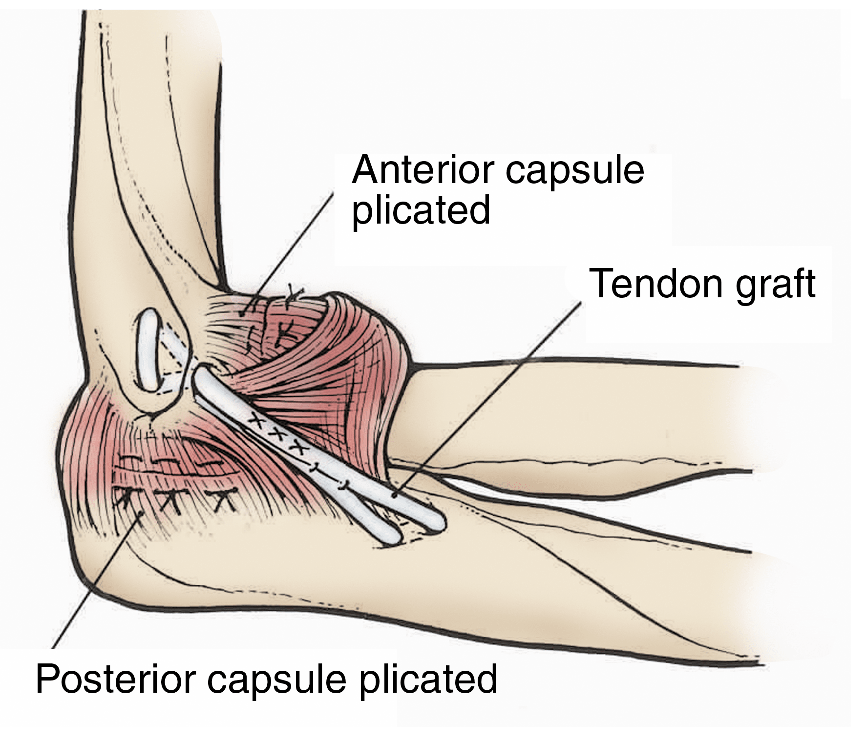
- Ligament reconstruction. Most ligament tears cannot be directly repaired or sutured (stitched) back together. To surgically fix the injury and restore elbow strength and stability, your doctor may need to reconstruct the ligament. During the procedure, the doctor replaces the torn ligament with a tissue graft, which serves as a new ligament. In most cases, the ligament can be reconstructed using one of the patient's own tendons (autograft), typically from the wrist or around the knee. However, sometimes the doctor will use an allograft (cadaver graft).
- Ligament repair. In some cases, when the ligament injury is relatively fresh or the remaining soft tissues are healthy, your surgeon may recommend repairing the ligament with sutures. The ligament repair can also be combined with an additional strong, thick suture, which acts as a belt or suspender to provide added support while the ligament heals.
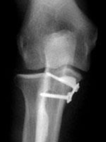
- Fracture fixation. Patients with unstable elbows with significant associated fractures require treatment to repair both the broken coronoid bone and the torn ligament. During the operation, the broken bone fragments are repositioned into normal alignment, then held together with special screws and, sometimes, a metal plate. If there are fractures of the radial head, those can be treated with similar fixation techniques, or the broken pieces can be replaced.
Recovery
During the first week after surgery, you will most likely wear a splint to protect your elbow as it begins healing.
Rehabilitation typically begins in the second week after surgery. The splint will be replaced with a brace that sometimes limits how far you can bend or straighten your elbow but allows you to begin exercises to improve range of motion while protecting the surgical fixation.
With a commitment to rehabilitation, you may regain full range of motion by 6 to 10 weeks after surgery. Given that both the type of instability and the combination of injured structures in the elbow will vary, your rehabilitation protocol will be customized for you by your surgeon.
Physical or occupational therapists will often prescribe strengthening exercises 3 months after the procedure, and most patients return to full activities by 6 to 12 months after surgery.
Throwing athletes may require up to 1 year of rehabilitation before returning to competitive sports.
Future Developments
Recurrent elbow instability is a relatively new concept. Future research will provide a better understanding of the interaction between the muscles, ligaments, and bones. Newer techniques are always evolving for reconstructing the ligaments. Research will lead to better ways to diagnose, treat, and recover from these complex injuries.
Contributed and/or Updated by
Peer-Reviewed by
AAOS does not endorse any treatments, procedures, products, or physicians referenced herein. This information is provided as an educational service and is not intended to serve as medical advice. Anyone seeking specific orthopaedic advice or assistance should consult his or her orthopaedic surgeon, or locate one in your area through the AAOS Find an Orthopaedist program on this website.


