


Diseases & Conditions
Aneurysmal Bone Cyst
Aneurysmal bone cysts (ABCs) are benign (not cancerous) lesions in bone, also called benign bone tumors.
Although ABCs are not cancer, they can be very destructive to bone and cause pain or pathologic fractures (breaks in the bone caused by a disease rather than an injury). They are more common in children than adults, typically appearing within the first 20 years of life.
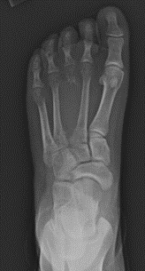
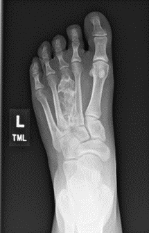
Aneurysmal bone cysts can appear in any bone in the body, but they are most commonly found in areas around the knee, shoulder, pelvis, and spine. They are usually discovered with an X-ray. They may be identified incidentally due to injury, or they may be identified due to pain and swelling in the affected area. Sometimes, the ABC is discovered when the bone breaks due to the cyst thinning and weakening the bone.
Some ABCs are more aggressive than others and can cause significant destruction of the bone.
Description
Aneurysmal bone cysts are rare skeletal tumors, affecting fewer than 1 in 100,000 people per year and accounting for 1 to 2% of all primary bone tumors.
- ABCs affect children more frequently than adults, with a slightly higher incidence in females.
- Most ABCs are hollow and are filled with different-sized sacs, called cysts. Each cyst has a lining, filled with blood and body fluid, that can change the shape and strength of the normal bone. This makes the bone more prone to fracture.
- A small percentage of ABCs are solid.
- Most ABCs are formed near growth plates (the areas at the ends of long bones in children and teens that allow the bones to grow longer)
- Most ABCs occur in a single bone.
While they may occur in any bone, ABCs are most commonly found in:
- The femur (thighbone)
- The tibia (shinbone)
- The humerus (the long bone in the arm that runs from the shoulder to the elbow)
- The spinal vertebrae (bones in the spine)

Cause
Doctors do not know what causes aneurysmal bone cysts. They are thought to occur as the bone is growing, as they are frequently found near the growth plates.
Sometimes, an ABC can develop in reaction to another benign bone tumor, such as a chondroblastoma, nonossifying fibroma, or giant cell tumor. This is called a secondary aneurysmal bone cyst, and it can be more challenging to diagnose.
Symptoms
- Patients can experience pain and swelling in a bone or joint. Often, pain starts without any injury or trauma. Pain or swelling may gradually develop and can get worse over time.
- Patients might have stiffness or decreased range of motion in the arm, leg, hand, or foot.
- Sometimes, a broken bone is the first sign that a person has an ABC. The person may feel a pop or a snap when they fracture the bone. Sometimes there is pain before the fracture occurs, but not always. The ABC is then discovered when the person goes to the emergency room to treat the fracture.
- If the ABC is in the spine, patients may experience back or neck pain, nerve pain radiating (spreading) into the hands or feet, or difficulty using the bathroom.
- Sometimes, there are no symptoms, and the ABC is discovered incidentally, such as during a routine doctor's visit or on an X-ray taken to confirm an injury.
Doctor Examination
Physical Examination
Your or your child's doctor will talk to you about your or your child's general health and medical history and ask about symptoms. They will examine the area that is concerning to look for:
- Swelling
- Stiffness
- Any signs of deformities
- Decreased range of motion
- Pain
- A mass
Imaging
X-rays. Your or your child's doctor will likely order X-rays to look at the underlying bone.
The X-ray typically shows a bone lesion that has changed the shape and the strength of the bone. Usually, the bone is enlarged with a clear space in the middle, and the bone cortex (strong outer layer) is thin.
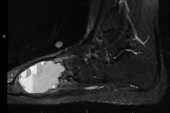
After reviewing the X-rays, the doctor may order magnetic resonance imaging (MRI) scans or, on very rare occasions, computed tomography (CT) scans.
Magnetic resonance imaging (MRI) scans. MRI scans help doctors determine where the tumor begins and where it ends, which helps them to plan treatment.
While CT scans are better at visualizing the bone, MRI scans are best for looking at the tissue and fluid inside the cyst. The classic appearance of an ABC on MRI shows fluid-fluid levels which represent blood and cyst fluid layered in the bone tumor.
Other Tests
Laboratory Tests. Most blood tests are not helpful in making a diagnosis of ABC.
Biopsy. A biopsy must be done to confirm the diagnosis of an ABC. A biopsy is a procedure used to get a tissue sample for a pathologist to analyze.
There are two ways to perform the biopsy:
- Core needle biopsy. The doctor places a small needle through your skin to get a small sample of the bone, which is then examined under a microscope.
- Open biopsy. If a larger sample is needed, the biopsy is performed in an operating room, and the doctor makes an incision to obtain the bone sample.
Genetic testing. A gene called USP6 has been found to be responsible for many aneurysmal bone cysts. The bone can be tested for the presence of USP6 by the pathologist as part of the biopsy to help make a diagnosis.
Treatment
There are many treatment options for bone and soft tissue tumors. Therefore, it is important to talk to your or your child's doctor, who will customize your treatment for the best possible outcome.
Surgery
Surgery is the mainstay of treatment for ABCs, and most patients undergo removal of the tumor. There are several surgical options:
Curettage and bone grafting
- The surgeon uses tools to scrape the tumor out of the bone. The goal is to remove as much of the tumor as possible without damaging the healthy bone or the growth plate.
- To help reduce the chance of the tumor returning, the surgeon may use a burr, a laser, hydrogen peroxide, liquid nitrogen, or phenol to try and destroy any microscopic tumor cells.
- After the tumor is removed, the surgeon will fill the empty hole in the bone with a bone graft or a bone graft substitute. Bone graft can be taken from the patient (autograft) or from a donor (allograft). Donor bone graft has all living cells removed and contains only the mineral structure of bone. It does not, therefore, require a special match or for the recipient to take medication to prevent rejection. Bone graft substitute is usually made of calcium phosphate or calcium sulfate with other minerals that help the body grow new bone.
- Sometimes, the surgeon needs to support the bone with plates and screws or other hardware.
- Sometimes, patients need to wear a splint, cast, sling, or brace while they recover.
Wide Resection
- Surgery with a wide resection, or en bloc resection, involves removing a large segment of bone where the cyst is located.
- Surgery with a wide resection is usually reserved for ABCs that have caused severe bone destruction or have recurred (come back) multiple times.
- Surgery with a wide resection usually requires the bone to be reconstructed or rebuilt with a large piece of donor bone (allograft) or a metal bone or joint replacement.
Sclerotherapy or Radiotherapy (Radiation Therapy)
Sclerotherapy and radiotherapy are saved for large, difficult-to-access tumors that would be challenging to remove safely during surgery.
Both treatments are commonly considered for ABCs in the spine, sacrum, or pelvis. For example, if you or your child have an ABC in the sacrum (the bone at the base of the spine), surgery could potentially damage important nerves that control the legs, bowel or bladder, and alternate therapies might be a safer option.
Sclerotherapy
This minimally invasive procedure involves directly injecting a solution into the tumor or blood vessels near the tumor. There are many different substances that can be injected, including polidocanol, doxycycline, or alcohol. This procedure allows patients to go home the same day, but multiple treatments are often needed to eliminate the tumor.
Radiotherapy
Radiotherapy uses a type of radiation to target the tumor cells. These high doses of radiation usually will kill the tumor and prevent it from coming back.
Recovery and Surveillance
The goal of treatment is to eliminate the bone cyst, improve pain, and allow the bone to grow more normally. Recovery time after surgery can vary based on the size and location of the ABC. It can take several months (usually 3 to 6 months) for the bone to heal.
Unfortunately, the bone tumor can come back. ABCs recur approximately 25% of the time. (This means the tumor grows back in 1 out of 4 patients). It is, therefore, important for you or your child to follow up with the surgeon as scheduled to check for appropriate healing and recovery.
After the bone heals, surgeons frequently start surveillance, a process of watching the bone with the tumor over time. The surgeon might monitor your or your child's ABC with X-rays every 3 to 6 months to make sure the tumor does not grow back.
While there is no set guideline, surveillance may continue for 2 years or until the patient reaches skeletal maturity (the bone is finished growing).
Contributed and/or Updated by
Peer-Reviewed by
AAOS does not endorse any treatments, procedures, products, or physicians referenced herein. This information is provided as an educational service and is not intended to serve as medical advice. Anyone seeking specific orthopaedic advice or assistance should consult his or her orthopaedic surgeon, or locate one in your area through the AAOS Find an Orthopaedist program on this website.